

A horse’s legs are a masterpiece of design. They carry incredible strength and delicate precision.
Every bone, joint, and tendon plays a role in their power and agility. Understanding the anatomy of these legs can help you care for them better.
From the sturdy cannon bone to the sensitive fetlock, each part is unique.
Whether you’re a rider, vet, or horse lover, knowing a horse leg anatomy can make a big difference.
Dive into the details and see what makes a horse’s legs both strong and fascinating.
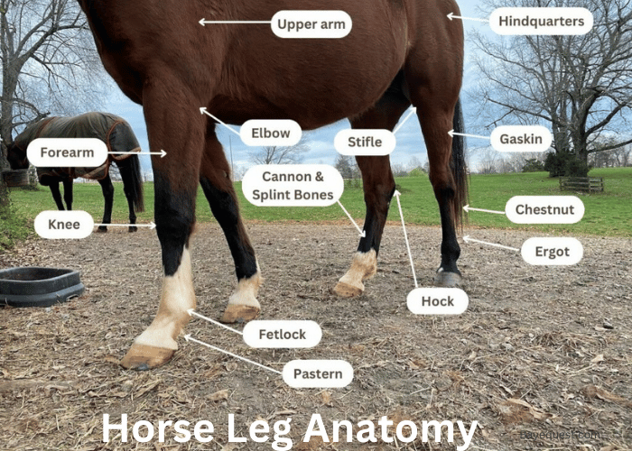
Front Leg Horse Anatomy
Let’s explore the anatomy of the horse’s front leg, understanding each part’s structure, location, and vital function.
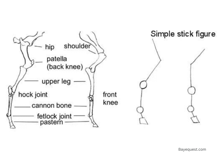
Shoulder
The horse’s shoulder is a sloping and muscular area at the top of the front leg. It connects the leg to the horse’s body.
The main bone here is the shoulder blade, or scapula. This area allows the horse a wide range of movement.
The slope of the shoulder affects the horse’s stride length and overall movement. A well-angled shoulder contributes to a smoother, more efficient gait.
Upper Arm (Humerus)
The upper arm, or humerus, is the bone that runs from the shoulder to the elbow. It is relatively short and thick.
This bone helps in transferring the body’s weight to the lower leg. The humerus provides leverage for the muscles that control leg movement.
Its strength and positioning are crucial for powerful strides and stability.
Elbow and Forearm
The elbow is the joint where the upper arm meets the forearm. It’s a prominent, bony area that you can easily feel.
The forearm consists of two long bones: the radius and ulna. This part of the leg extends from the elbow to the knee.
The elbow joint allows flexion and extension, while the forearm provides support and strength for movement.
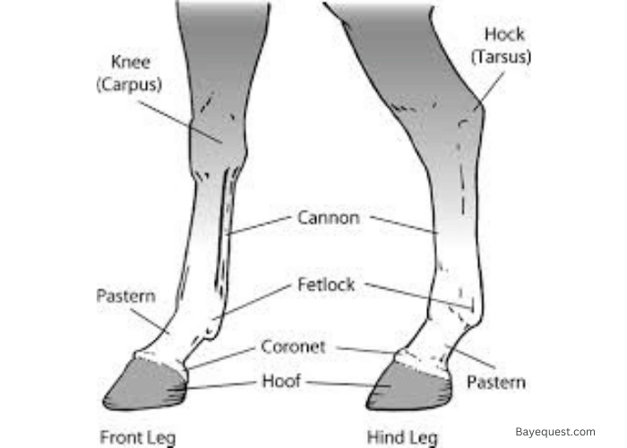
Knee (Carpus)
The knee, or carpus, is a complex joint midway down the front leg. It consists of several small bones and acts like the human wrist.
This joint allows the horse to bend and absorb shock. The knee is crucial for the horse’s ability to move quickly and change direction.
It’s also a common site for injuries due to its complexity and the stress it endures.
Cannon Bone and Fetlock
The cannon bone is the large, strong bone below the knee. It runs down to the fetlock joint.
The fetlock is a flexible joint that allows the leg to move and absorb impact. This area is essential for weight-bearing and shock absorption.
The cannon bone provides strength, while the fetlock’s mobility is key to a smooth gait.
Pastern and Hoof
The pastern is the area between the fetlock and the hoof. It consists of two small bones and is angled slightly.
This angle helps absorb shock and contributes to the horse’s smooth movement. The hoof is the hard outer part of the foot.
It protects the internal structures and bears the horse’s weight. Proper hoof care is vital for a horse’s overall health and performance.
Sesamoid Bone
The sesamoid bones are two small bones at the back of the fetlock joint. They act as pulleys for tendons and provide leverage.
These bones play a key role in the movement and flexion of the leg. They help reduce friction and protect tendons from excessive wear.
Damage to these bones can significantly affect a horse’s mobility.
Pedal Bone
The pedal bone, or coffin bone, is located inside the hoof. It is the main bone within the hoof and shapes the hoof itself.
This bone supports the horse’s weight and provides attachment points for various tendons. It’s crucial for the horse’s stability and movement.
Any issues with the pedal bone can lead to serious lameness.
Navicular Bone
The navicular bone is a small, boat-shaped bone located behind the pedal bone and below the sesamoid bones. It acts as a fulcrum for the deep digital flexor tendon.
This bone helps distribute pressure and reduce friction within the hoof. Problems with the navicular bone can lead to chronic lameness known as navicular syndrome.
Proper care and management are essential to prevent issues in this area.
Rear Leg Horse Anatomy
Let’s delve into the anatomy of the horse’s rear leg, exploring each part’s structure, location, and essential function.
Hip and Pelvis
The hip and pelvis are located at the top of the horse’s rear leg. The pelvis is a broad, sturdy bone structure that supports the horse’s hindquarters.
The hip joint connects the pelvis to the femur. This area provides strength and stability, crucial for powerful movements like jumping and galloping.
Femur and Stifle Joint
The femur is the long bone running from the hip to the stifle joint. The stifle joint is similar to the human knee. It allows the leg to bend and extend.
This joint plays a key role in the horse’s ability to move forward and is vital for proper locomotion.
Read more on stifle joint injury in our guide.
Gaskin (Tibia and Fibula)
The gaskin is the area between the stifle and the hock, containing the tibia and fibula. These long bones provide leverage and support.
They are essential for transmitting the force the muscles generate to the lower leg, enabling powerful strides.
Hock Joint
The hock joint is a complex joint located midway down the rear leg. It acts like the human ankle and comprises several small bones.
The hock allows for a wide range of motion and absorbs shock. It is crucial for the horse’s ability to jump, kick, and move quickly.
Cannon Bone and Fetlock
The cannon bone is a strong bone below the hock that runs down to the fetlock joint.
The fetlock is a flexible joint that supports the horse’s weight and provides shock absorption.
This combination of strength and flexibility is key for smooth, powerful movement.
Pastern and Hoof
The pastern is the area between the fetlock and the hoof, consisting of two small bones. It helps absorb shock and contributes to the horse’s movement.
The hoof is the hard outer structure that protects the internal parts and bears the horse’s weight. Proper hoof care is essential for the horse’s health.
Patella (Kneecap)
The patella, or kneecap, is a small bone in front of the stifle joint. It helps protect the joint and provides leverage for the muscles controlling leg movement.
The patella is crucial for the stability and function of the stifle joint, aiding in the horse’s overall mobility.
Importance of Healthy Horse Legs
Healthy horse legs are crucial for a horse’s overall well-being.
Strong legs mean better movement and performance. They allow the horse to run, jump, and work without pain.
Healthy legs support the horse’s weight and absorb shock. This prevents injuries and long-term problems.
Regular care and check-ups can spot issues early. Proper shoeing and a good diet also play a big role.
If a horse’s legs are healthy, the whole horse is healthier and happier. It’s that simple.
Taking care of their legs is one of the best ways to ensure your horse stays active and strong.
Common Horse Leg Injuries and Conditions that Affect the Equine Leg Anatomy
Let’s explore common leg injuries and conditions in horses, their symptoms, causes, and how to manage them effectively.
Lameness
Lameness means a horse is limping or moving unevenly. It’s often caused by pain in the legs.
This can come from various issues like arthritis, abscesses, or injuries. If a horse is lame, you need to find the cause quickly.
Tendonitis
Tendonitis is inflammation of the tendons. These are the bands of tissue that connect muscles to bones.
It often happens from overuse or sudden strain. Signs include swelling, heat, and pain in the affected area.
Laminitis
Laminitis is a serious condition affecting the hooves. It causes inflammation of the tissue inside the hoof.
This can be extremely painful and may lead to permanent damage. Common causes include overeating, infections, or trauma.
Hoof Abscesses
Hoof abscesses are pockets of infection inside the hoof. They cause severe pain and sudden lameness.
Abscesses usually occur from a puncture wound or a crack in the hoof. They often need to be drained and treated with antibiotics.
Navicular Disease
Navicular disease affects the navicular bone and surrounding tissues in the hoof. It causes chronic pain and lameness.
The exact cause is often unknown but can be related to poor hoof conformation or hard work on rough surfaces.
Arthritis
Arthritis is the inflammation of the joints. It causes pain, stiffness, and swelling.
It’s common in older horses and those with heavy workloads. Managing arthritis often involves pain relief and careful exercise routines.
Related read: How to Treat Arthritis in Horses.
Fractures
Fractures are breaks in the bone. They can be caused by falls, kicks, or collisions.
Fractures need immediate veterinary attention and often require long rest periods to heal properly.
Suspensory Ligament Injuries
The suspensory ligament supports the horse’s leg during movement. Injuries here can cause lameness and swelling.
These injuries are common in performance horses due to the strain of intense activity.
How to Treat Horse Leg Injuries
Treating horse leg injuries requires patience and proper care. Follow these steps to help your horse recover and get back to its healthy, active self:
1. Rest
First and foremost, rest is crucial. If your horse is injured, give them time off. Don’t let them run or work. Rest helps the healing process and prevents further damage.
2. Cold therapy.
Use cold therapy to reduce swelling and pain. Apply ice packs or cold water to the injured area. Do this several times a day for 15 to 20 minutes each time.
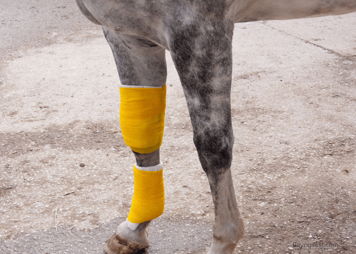
3. Compression.
Compression can help control swelling. Use bandages to wrap the injured leg. Make sure they’re snug but not too tight. You don’t want to cut off circulation.
4. Elevation.
If possible, elevate the injured leg. This can help reduce swelling. However, this is more practical for minor injuries or when the horse is lying down.
5. Medication.
Your vet might prescribe anti-inflammatory drugs. These can reduce pain and swelling. Always follow the vet’s instructions carefully when giving medication.
6. Physical therapy.
Once the initial swelling goes down, your horse might benefit from physical therapy. This could include gentle stretching and controlled exercise. It helps restore strength and flexibility.
7. Proper diet.
A healthy diet can aid in recovery. Ensure your horse gets enough nutrients, especially those that support joint and bone health. Consult your vet for dietary recommendations.
8. Regular check-ups.
Keep in touch with your vet. Regular check-ups can ensure the injury is healing properly. Your vet can also adjust treatment plans as needed.
9. Avoiding recurrence.
Finally, prevent future injuries by maintaining proper hoof care, using the right shoes, and ensuring your horse has a safe environment. Avoid overworking your horse and gradually increase their workload.
Related read: Horse Muscle Anatomy.
Anatomy of Horse Leg: FAQs
What is the lower part of a horse’s leg called?
The lower part of a horse’s leg is called the cannon bone, which extends down to the fetlock, pastern, and hoof.
What are the ligaments in the lower leg of a horse?
The ligaments in the lower leg of a horse include the suspensory ligament, the check ligaments, and the digital flexor tendons’ supporting ligaments. These ligaments provide stability and support for movement.
Horse Leg Anatomy: Conclusion
Understanding a horse’s anatomy isn’t just for vets. It’s for anyone who loves and cares for horses.
Knowing the ins and outs of their legs helps keep them happy, healthy, and ready to gallop.
From the sturdy shoulder to the hardworking hoof, every part plays a role.
So, next time you watch your horse run, you’ll appreciate the marvel of their legs even more.
Keep learning, keep caring, and your horse will thank you with every graceful stride.